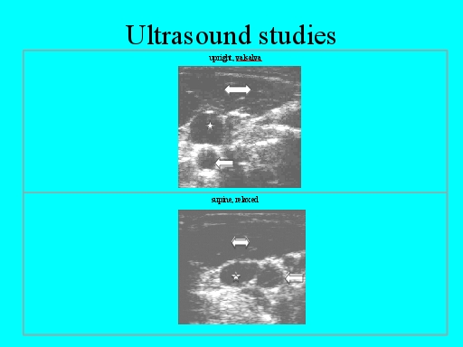
Here’s my own internal jugular vein. Upper photo standing with valsalva, note the position of the internal jugular vein between the SCM and IJ, Zone I. Legend: double arrow- sternocleidomastoid muscle, left arrow- carotid artery, star- internal jugular vein.
Lower photo lying down, no valsalva, look at the large area of the IJ, comparable to a standing valsalva, but remember that in a standing position, the IJ is almost invisible due to compression.
Previous studies have described this IJ change in a supine position as a shape change allowing increased blood flow. In reality, in my studies, it was discovered, as you can see from careful examination, that the internal jugular vein has fallen laterally out of Zone I compression. This occurs because as the body becomes supine, the vein begins to fall laterally into the loose surrounding tissue, fills with blood, which then, due to the weight of the blood in the vein, pulls the IJ even farther out of Zone I as it falls laterally. This falling out of Zone I does not occur in microgravity because, of course, there is no weight to cause the IJ to fall. KEY POINT.